|
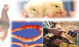 Coccidiosis in one of the major menace for poultry industry
causes heavy economic losses world wide. The disease is
commonly called as Red Dysentry (Khuni pachish). Coccidiosis
may be Intestinal or caecal caused by intracellular protozoa,
belonging to the genus Eimeria (Phylum Apicomplexa). Coccidiosis in one of the major menace for poultry industry
causes heavy economic losses world wide. The disease is
commonly called as Red Dysentry (Khuni pachish). Coccidiosis
may be Intestinal or caecal caused by intracellular protozoa,
belonging to the genus Eimeria (Phylum Apicomplexa).
The
disease is characterized by bloody diarrhoea, emaciation,
ruffled feather and loss of apatite. More or less about 300
species of genus Eimeria has been known and recorded in birds
and mammals. It is estimated that the economic losses due to
the disease are about US$ 450 million and due to medication
are about US$ 100 million in the United States.
In countries
like Pakistan, where the farming is substandard, the disease
becomes more serious and causes heavy economic losses;
although the exact losses due to coccidiosis in Pakistan are
not known due to the lack of statistical indices but these
will be definite in million of rupees.
Seven species of genus Eimeria (E.) including E. tenella, E.
acervulina, E. maxima, E. necatrix, E. mitis, E. praecox and
E. brunetti are generally accepted to be the causative agent
of avian coccidiosis. E. tenella and E. maxima are considered
to be the most important to the poultry industry from
consideration of their ubiquity in broiler chicks, innate
pathogenicity and immunological features. In Pakistan E.
tenella is the most prevalent and pathogenic species.
The conventional methods to control the disease are by using
certain coccidiostats/coccidiocidal drugs, but
in-discretionary use of anticoccidial drugs including
monensin, amprolium, ionophores and nicarbazin has resulted in
the emergence of drug resistant strains, which has reduced the
efficacy of many of the currently used coccidiostats. During
the recent years, pharmaceutical industries throughout the
world have not marketed any new anticoccidials. Obviously an
alternative system to control the disease is by vaccination.
Time to time different attempts have been made to immunize the
chicks against coccidiosis throughout the world by using
different antigenic materials including low doses of
sporulated oocysts , irradiated sporulated oocysts,
sporozoites, merozoites, recombinant vaccine, recombinant
refractile body antigen and inactivated sporulated oocysts.
But none of the experimental vaccine reached to commercial
scale up to 1980. In late 80’s and early 90’s, Immnocox,
Paracox and Livacox was developed and are being marketed now
in different countries including Canada, Bangladesh,
Scandinavian countries, India, Pakistan and European
countries. Immunocox is also available commercially in
Pakistan although it is not used routinely in the commercial
flocks due to disease occurrence even in the vaccinated birds;
the vaccine failure may be due to the strain variations.
Keeping in view, an experimental sporulated vaccine from local
isolates of coccidia was prepared in the Immunoparasitology
laboratory, Department of Veterinary Parasitology, University
of Agriculture, Faisalabad gave promising results but it was
not to be commercialized due to the non-availability of
antigen in large scale. It was, therefore, imperative to adopt
the strategies that lead to the production of antigenic
material on large scale to be used in vaccine preparation.
Keeping in view a study was planned having the following
objectives:
a. Adaptation of E. tenella (local isolate) on the
chicken embryos.
b. Formulation of vaccine(s) from egg-adapted
gametocytes.
c. Evaluation of vaccine(s) on the basis of, mucosal,
cellular, humoral and challenge responses.
Keeping in view, successful attempts were made in the
adaptation of E. tenella, the most pathogenic species, on the
chicken embryos.Life cycle of E. tenella sporozoites was
established on the chicken embryos on chorio-allantoic
membrane. It completed its life cycle in 6th-7th days.
Sporozoites were injected into chorio-allantoic membrane of
ten-days-old chicken embryo. Mature and immature schizonts
were seen in CAM on day 4, 5, 6 and 7, and numerous merozoites
were observed in the chorio-allantoic fluid. Gametocytes and
Oocysts were also found in Chorio-allantoic fluid (CAF).
Gametocytes and oocysts were detected in CAF 6th to 7th days
post inoculation of sporozoites. Gametocytes (macro &
microgamets) were obtained in the CAF. During that period, 2%
death in embryo was observed from day 1 to 4th that may be not
associated with parasitaemia and due to the toxic substances
From day 5th to 7th, 60.8 % embryos were found dead and 34.4 %
embryos were found alive. Enormous numbers of gametocytes were
produced after the inoculation of sporozoites into the CAM.
These egg adapted gametocytes were used in the vaccine(s)
formulation viz vaccine-I (gametocytes), vaccine-II
(gametocytes inactivated with formalin) and vaccine-III
(inactivated sonicated gametocytes) and were given orally to
immunize the chicks.
The gameyocytes vaccine(s) were evaluated on the basis of
cellular, humoral, mucosal and challenge responses. All the
vaccine(s) gave protection against heavy doses of infection of
mixed species of coccidia.
Cell mediated immune response of the vaccinated and control
Groups was detected by splenic cell migration inhibition assay
and results were expressed interms of migration index
calculated from cellular migration distance from the edge of
fragment with and without antigen. On day 5th and 15th post
vaccination, there was significant difference (P<0.01) in
migration index of all four Groups with antigen and
non-significant difference without antigen. This indicates
that the test antigen has triggered the T-cells; to initiate
the cell mediated immune response.
Humoral response in chicks vaccinated with gametocytes
vaccine(s) was detected by indirect haemagglutination test and
results were expressed interms of geomean antibody titre. A
non-significant difference (P>0.05) in geomean titer among
Vaccine I, Vaccine II, Vaccine III and control Group at day
15th post vaccination was recorded. On the other hand, the
difference (P<0.01) in geomean titer among Vaccine I, Vaccine
II, Vaccine III and control Group was significant on day 21st
post vaccination.
IgA, IgG and IgM play vital role in the binding of foreign
antigens and the presence of these antibody molecules on a
microbial or parasitic surface can cause clumping
(agglutination) and among them IgG activates the complement
system.
In the present study, Enzyme linked immunospot (ELISPOT) assay
was used to detect the IgA, IgG and IgM antibodies producing
cells in chicks vaccinated with gametocytes vaccine(s) against
coccidiosis.
There was significant difference (P>0.05) of IgA antibody
secreting cells of Group III with Group II and I and
non-significant difference (P>0.05) between Group I and II.
Significant difference (P>0.05) of IgG antibody secreting
cells of Group III with Group I and II and non-significant
difference (P>0.05) between Group I and II was recorded.
Similarly significant difference (P>0.05) was also recorded of
IgM antibody secreting cells of Group III with Group I and
Group II and non-significant difference (P>0.05) between Group
I and II. Results of the present study revealed that spleen is
one of the major sources of cells producing IgA, IgG and IgM
secretors. IgA and IgM antibody secreting cells were maximum
in Group III followed by Group II and Group I. IgG antibody
secreting cells were also maximum in Group III but numbers
were equal in Group II and Group I. Such a high level of
immunoglobulins secretors in Group III (inoculated with
sonicated formalized gametocytes vaccine) indicate the
synthesizes of Major immunoglobulins particularly IgA and IgG
that has a major role to prevent adherence of coccidial
parasite to the body of the host. From the ELISPOT assay
results it may be concluded that Vaccine III gave better
protection followed by Vaccine II and Vaccine I.
Challenge experiments were also conducted to evaluate the
gametcyte vaccine(s). For this purpose, oocysts of mixed
species of genus Eimeria were used to challenge the vaccinated
and control birds. There was non-significant difference
(P>0.05) in oocysts count between Group I and Group II while
the difference of Group III was significant (P>0.05) with
Groups I, Group II and Group IV. Chicks mortality in Group I
was significantly higher (P>0.05) as compared to Group III and
control Group IV but mortality difference was non significant
different (P>0.05) between Group-I and Group II. Results of
the challenge experiments showed that the Group III gave
vaccine III resisted to heavy dose of infection. There was
non-significant difference (P>0.05) in lesions score of Group
I, Group II and Group IV.
Lesions scores in Group III were significantly different
(P>0.05) from Group I, Group II and Group IV. Lesion scores
were found to be directly proportional to the mortality and
oocyst count per gram of faeces/droppings but inversely
proportional to the percent protection and level of the immune
responses. It was also observed that mild to moderate lesions
produced despite of immunity conferred which reflect that the
organism has multiplied but may not lead to fatality to cause
the disease or mortality.
On the basis of cellular, humoral, mucosal and challenge
responses, Vaccine III (inactivated sonicated gametocytes) was
found to give better results as compared to Vaccine II
(gametocytes inactivated with formalin) and Vaccine I
(gametocytes).
Further studies on the molecular characterization of egg
adapted gametocytes antigen to identify the candidate antigen
and its feasibility on commercial scale.
PAKISSAN
|
Pakissan.com;
|