|
MASTITIS: A Monster Threat to Dairy
Industry
By
definition Mastitis is “inflammation of the mammary gland”.
 In
the present state of knowledge it seems practicable and
reasonable to define mastitis as a disease characte- rized
by the presence of a significantly increased leukocyte count
in milk from affected glands. In
the present state of knowledge it seems practicable and
reasonable to define mastitis as a disease characte- rized
by the presence of a significantly increased leukocyte count
in milk from affected glands.
The Mastitis Committee of
the Australian Veterinary Association defined nature and
causes of mastitis as “Mastitis is an inflammation of udder
and as such is a disease complex resulting from any
condition or combination of factors leading to injury to the
internal structure of one or more quarters”.
There is no standard
definition of various types of mastitis but is simplest to
consider mastitis as clinical or sub-clinical&symptoms may
be acute or chronic.
Clinical Mastitis:
This is characterized by heat, pain and swelling or in
duration, which may occur with or without these signs. The
udder secretion is usually abnormal; milk yield and quality
are usually markedly affected. Several factors influence the
severity of the effects e.g., organism responsible, the
susceptibility of the cow and the extent of udder damage.
Sub-clinical Mastitis:
A quarter infected with a pathogen, having an increase in
the cell content of the milk and the absence of clinical
signs, is generally accepted as being affected with
sub-clinical mastitis. Indirect tests such as California
Mastitis Test (Rapid Mastitis Test in Australia), cell count
or white side test are required to make the diagnosis. The
level of cell count regarded as significant varies with
different workers, but counts in excess of 500,000 cells per
ml are generally regarded as indicative of sub clinical
mastitis. This form of mastitis is frequently not noticed by
the farmer.
Latent Infection:
This refers to infection with mastitis organisms, which has
not produced changes coming within the deficiency of sub
clinical mastitis. This condition is most common with
staphylococci and streptococci. Most potentially pathogenic
bacteria have been implicated at one time or another in
causing mastitis, as have yeasts and Mycoplasma organisms.
However, Str. agalactiae, staph aureus, other streptococci
and Gram-negative bacilli (including E. coli and Pseudomonas
spp. etc.) account for probably 99% of all mastitis. It is
clear that the first two infections are the more important
and probably staphylococci are now the commonest organisms
associated with mastitis.
Mastitis is a disease of all milking animals. It is common
in dairy cattle, uncommon in beef cattle. In cattle it is
characterized by changes in the udder tissue, clots and
changes in the constitution of milk, and is sometimes
accompanied by heat and pain in the udder.
Causative Agents:
When we think of the extreme complexity and delicacy of the
mammary gland, the fact that it is pummeled along between
the hind legs of the cow, lain on, kicked, horned; that it
contains milk, a suitable medium for the growth of a great
number of bacteria, fungi and other micro-organisms; that
such milk is open, via the teat canal to contamination and
is interfered with at least twice a day, it is surprising
that any cow ever escapes mastitis.
Added to this, by
selective breeding the gland has been developed to
an abnormal size and selected to secrete a grossly
abnormal amount of a natural product.
It is thus apparent that
particular efforts must be made to treat the udder with
every possible care to eliminate predisposing causes which
include, the presence of chronically affected carriers in
the herd, the use of dirty methods of milking which
transfers infectious milk from animal to animal, and faulty
milking machines which may by excessive suction, cause
damage. Faulty sphincter of the teat may give imperfect
closure of the teat canal and facilitate infection. Sores at
the opening of the teat canal probably play an important
part in allowing the germ to enter. Infection with
contagious abortion, and also an inherited predisposition
are predisposing causes mentioned by some authorities.
Feeding a high protein grain ration apparently renders the
udder more liable to infection. Chronically, infected udders
are liable to “Flare up” into the acute form. Faulty
milking, filthy conditions and the presence of flies may all
play a part in assisting the spread of the disease.
Mechanical injury of the udder will, of course, act as a
definite predisposing cause.
It is important to realize
that milk is secreted, in part, by erectile tissue in the
udder becoming turgid with blood. Unless the cow is settled
and contended and enjoys milking time, it does not occur.
Such reaction is a conditioned reflex. Thus, if a cow is
accustomed to being washed with water, say one minute before
she is milked, her ductless glands will so secrete their
hormones that at this given time after the washing with warm
water the erectile tissue will become turgid and the milk
will flow into the teats. As the farmers say “she lets down
her milk”. This “letting down” the milk is conditioned in
response to all the usual happenings. These happenings
should be so masterminded that each event becomes a
pleasuring device. The wise farmer “woos” fickle female
bovines to yield up all their milk by “pleasuring” the
erectile udder tissue to climax and so “let down” the milk.
Thus, if the cow has her
udder washed at a longer period than usual before milking is
commenced or if she comes into the bail (stanchion or tying
place) out of order, or is forced to go into the wrong bail
or is otherwise upset (e.g. by the presence of dogs or
strangers, kicks or horning), the secretion of milk may be
interfered with. Under such circumstances the udder is not
milked out and this may have a major effect in precipitating
outbreaks of mastitis. In practice the employment of a new
milking hand or some sharp change in procedure is often
noticed to coincide with the occurrence of mastitis.
Music in bails has been found
to have a considerable effect on the contentment and milk
secretion of the cows. Cows particularly some nervous and
highly-strung breeds, accustom themselves to particular
persons. It is well known that some attendants with a good
“animal sense” have a vast influence on the contentment and
production of the animals they handle. In control of
mastitis this factor of the wise contended handling of cows
is of the very greatest importance.
The features of chronic bovine mastitis were closely
resembled with the features of tissue allograft undergoing
rejection. Workers further showed that milk is rich in
leukocytes and, if milk from one cow is injected into the
quarter of another cow previously milked out, the foreign
leukocytes stimulate an allogenic lymphocyte transfer
reaction. They further pointed out that a pipeline milking
machine might transfer milk from one cow’s udder into the
empty udder of another cow when it is incorrectly managed.
They consider that in some cases this may be an important
predisposing cause to clinical mastitis in a cowshed.
The incidence of heifers shedding “staphylococci” in the
milk increased from very low levels to approximately 50% by
8 weeks. When heifers were milked separately from older
cows, through teat cups that were chemically sterilized
between milking, under these conditions the number of
heifers shedding “staphylococci” during their first
lactation was on average 10% or less.
In a complex disease such as mastitis, predisposing factors
are manifold. Smith et al. (1984) showed that selenium
deficiency is associated with increased susceptibility to
mastitis. Other workers showed that it is also associated in
cattle with retained fetal membranes, ill thrift,
reproductive disorders and reduced response to some
bacterial infections. Thus, in selenium deficient areas the
provision of proper dose of selenium will helpful in
preventing the mastitis. Ryan et al. (1987) produced good
controlled evidence that selenium deficiency predisposes to
staphylococcal mastitis infection. It has been shown that
pastures high in estrogens may predispose cows to mastitis.
Infectivity
a. Susceptible Animals
All breeds of dairy cows are susceptible. It is generally
held that high producers are more commonly affected than
poor producers. With succeeding lactations, the liability to
have contracted infection increases. That is, more cows on
fourth calf than cows on their first calf are affected.
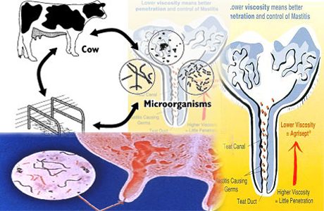
b. Mode of Infection
All modes of infection may operate. The causal bacteria may
be transferred by the hands of the milkier, by milking
machine cups and by flies, or the cow may be infected
directly by lying on contaminated ground. Infection may be
up the teat canal, through the blood stream or through skin
injuries. Apparently, the mode of infection tends to vary
with each bacterial type. In some cases it has been shown
that the organisms grow up the teat canal, cause
inflammation of the milk cistern at the base of the teat and
then spread to other parts of the gland, causing
inflammation of the whole quarter. In case of “streptococcus
agalactiae”, the main mode of infection is via teat canal,
and this fact lies behind nearly all the hygienic methods of
control recommended.
c. Immunity
Cows that have been affected or are recovering from one
attack do not remain immune to further attacks.
Signs And Symptoms:
A wide range of differing symptoms will be shown with
different types of infection. Milk may become watery or
thickened or pus-like, and may contain clots which vary in
color from yellow to blood clots. The clots may also vary
considerably in character. In some infections they are
large, slimy clots. Other clots may be purulent; some
infections produce mealy or flaky clots. Some clots may
consist of fine flocculation that is seen like small pieces
of blotting paper. The nature of the milk may be changed in
all sorts of ways. Thus, on some occasions, it is merely
thinner and paler. It may be thickened and amber in color,
darkened, or blood stained, and have varying degrees of
unpleasant smell. Milk production may be decreased or ceased
altogether and be replaced by pus, clots or watery material.
The condition of the udder may become hot, tense, and
painful and later develop fibrotic changes with hard lumps,
or it may be shriveled and wasted. In some severe types, one
or more quarters of udder may die and slough right off
leaving a filthy, gaping wound. In severe cases the animals
will show general symptoms of toxemia or septicemia, being
off their feed, shivering and scouring and may die.
Management And Mastitis Spread:
Decreasing Exposure to Pathogen between Milking
The microorganisms are abundant in the surroundings in which
cows live, including manure, soil, bedding, feedstuffs,
water and plant material. Where poor hygiene exists, housed
cows are at greater risk of infection with environmental
microorganisms than cows on grassy pasture. However, where
good hygiene practices exist, housed cows may be at less
risk than cows on pasture that have access to ponds, mud
holes, or wet lots. The prevalence of clinical cases
increases with confinement; especially during winter months.
Environmental conditions that can increase exposure to these
microorganisms include overcrowding, poor ventilation,
inadequate manure removal, poorly maintained free stalls,
access to farm ponds, dirty calving areas, and general lack
of farm cleanliness and sanitation. Maximum air movement
through housing, feeding and calving facilities should be
provided to reduce the number of these microorganisms;
especially airflow over bedding materials, floors, and
walkways. Moisture of any kind such as rain, humidity,
urine, drinking water and even udder wash favor the growth
of environmental microorganisms.
Recommended Bedding Materials:
Low-moisture inorganic materials such as limestone, sand and
clay are preferable to finely chopped organic materials
because they contain few nutrients for bacteria to utilize
and harbor and are therefore associated with lower numbers
of microorganisms. Of these, washed sand is often
recommended because of greater ease in handling and because
it is less likely to harden in stalls if the material
becomes wet.
The key to successful use of any of the materials is daily
replacement of wet and soiled bedding and regardless of the
material used, this practice has been shown to reduce
bacteria counts. Composting solid wastes for bedding
material has been attempted, but it contains excessive
numbers of coliform bacteria once it is placed into free
stalls. Alternative bedding that has been evaluated is
recycling newspaper, but little advantage was gained in
decreasing bacterial counts which are similar to those in
chopped straw. However, it may be an economical alternative.
Prevention of Mastitis by Good Milking Procedures
1. Provide cows with a clean, stress-free environment
• Milking time should be a consistent routine.
• The cow should not be frightened or excited before milking
because such stress results in secretion of epinephrine in
blood stream which interferes with normal milk let down.
• Udders should be clipped or singed as necessary to remove
long hair and reduce amount of dirt, manure and bedding that
may contaminate milk.
• Hands of person milking the cows be washed and dried
before milking. Gloves are also beneficial. Hands/gloves be
rinsed in disinfectant solution before and after milking
each cow in a herd.
2. Check the foremilk and udder for mastitis
• Strip cups and plates should be cleaned and sanitized
after each milking to prevent the spread of mastitis
organisms.
• Stripping of milk directly onto the floor, followed by
immediate hosing of the floor surface can be done and
incorporation of black tiles into the floor of parlor
facilitates this procedure.
• Milk should never be stripped directly into the milker’s
hand because this procedure spreads microorganisms from teat
to teat and cow to cow via contaminated hands.
3. Wash teats and the Ventral surface of the udder with a
warm sanitizing solution
• Correct washing and massaging of teats and udder sends a
signal to pituitary gland that secretes oxytocin that causes
milk let down.
• In stanchion barns a sanitizing solution should be used in
bucket with individual cloth or paper towel to wash the
teats.
• Use of a common cloth or sponge should not be permitted
because these become grossly contaminated and may cause
mastitis spread.
In large dairy herds, milking facilities may be equipped
with sprinkler pens where 80 to 120 cows are group washed.
After providing them sufficient drip-drying time before cows
enter the milking parlor their teats and udders be
completely dried.
4. Use a pre-milking Teat Dip
Pre-milking teat dip reduces infections with environmental
microorganism by about 50%. Recommended pre-dipping
procedures are as follows: clean teats, forestrip predip
teats and allow recommended contact time (usually 20-30
seconds), dry teats with an individual paper towel to remove
germicide residues and attach milking units.
Pre-dipping is sometimes done without prior washing of the
dirty teats, and germicide is often placed on top of manure
and dirt present on teat skin. This practice is not likely
to reduce incidence of mastitis and somatic cell count, and
will probably reduce milk quality. Manure and dirt must be
removed to get full benefits of predipping.
5. Dry teats thoroughly
The milking of wet teats also promotes squawking of the teat
cup liners, which may result in teat end impact.
6. Attach teat-cups within 1 minute
Attachment of teat cups should be done carefully to prevent
the entrance of excessive air into the milking system.
Maximum internal udder pressure is reached approx. 1 minute
after udder preparation is begun and lasts for about 5 min.
Since the majority of cows will milk out in 4 to 6 min, the
consistent attachment of teat cups 1 minute after the
beginning of stimulation makes maximum use of milk let-down
hormone oxytocin.
7. Adjust milking units as necessary
• Teat cups that are seated excessively high on teats cause
irritation to the lining of the teat and may contribute to
the development of mastitis. Improperly aligned milking
units may also block milk flow, increase strippings and slip
more often.
• It is important that slipping or squawking of teat cups be
minimized because such occurrences probably contribute to
more machine-induced infections than any other single
factor.
• If liner slips occur at the same time as the liner opens,
tiny droplets of milk may be propelled against the end of
the teat at very high velocity. Such droplets may contain
mastitis-causing microorganisms and may penetrate the teat
canal. Since milk flow slows near the end of milking the
chances of the microorganisms being flushed out of the teat
are reduced and an infection of the quarter may result.
8. Shut off vacuum before removing teat cups
The goal should be to remove teat cups just as the last
quarter milks out, but vacuum should always be shut off
before teat cups are removed. An increased risk of infection
exists when teat cups are removed while under vacuum.
Dip teats with a safe and effective teat dip:
Teat dips in addition to killing all microorganisms on teats
reduce teat canal colonization and help to heal teat cup
lesions. The list of teat-dip germicides includes iodophores,
chlorohexidines, linear dodecyle benzene sulfonic acid (LDBSA),
sodium hypochlorite, sodium chlorite/lactic or mandelic
acid, hypochlorous acid, quaternary ammonium and
antimicrobial proteins and fatty acids.
Only products shown by research to be safe and effective
should be used. This involves using a product registered
with the FDA or concerned regulatory authorities.
Problems related to teat dips:
Some teat dip germicides may cause harmful effects on teat
skin and cause chapping. The irritation might be due to low
or high pH or high titratable acidity or alkalinity or a
formulation. Because of the potential for irradiation,
skin-conditioning agents often are added to teat-dope
formulations. Glycerin is an example of humectants, a
substance that promotes the retention of moisture.
Emollients, substances that soften and smooth the skin also
are found in dips; linolin is a popular choice and coats the
skin and reduces evaporative water loss. The only difficulty
is that germicidal activity of teat dips may be reduced if
concentrations of humectants and emollients become too high
above 10 or 12% of the total dip.
Normal teat skin is coated with bacteria static acids that
retard the bacterial growth. When exposed to cold, wet and
windy conditions, teat skin may become chapped and
irritated. Also protective surface coating may be removed,
allowing bacterial growth on teat skin.
Machine Milking
Mastitis spread/management
Basically, if equipment is operating according to the
recommendations, and its capacity is not exceeded, the
machine itself contributes little to the mastitis problem.
However, the milking system can influence the development of
mastitis causing bacteria from one cow to the next. Improper
use, such as failing to shut off vacuum when teat cups are
removed, may injure the teat canal and increase
susceptibility to infection. Irregular fluctuations in
milking vacuum may cause tiny droplets of bacteria-laden
milk to impact against the end of the teat, propelling
mastitis-causing bacteria through the teat opening and into
the udder. Therefore, it is important to dry teats before
milking, position the milking unit properly on udder, select
appropriate liners, avoid excessive machine stripping, and
minimize sudden air losses when machines are removed. Some
experts claim that changing liners every 1000 to 1200
milking or every 60 days, whichever comes first will reduce
risk of new infection.
Economic Importance
The losses caused to the dairy industry by this disease are
enormous. Almost every herd suffers intermittent losses from
good cows going ‘light’ or going blind in various quarters.
The aggregate loss to the industry is one of the major
deductions from economic production. It is probable that in
some herds more than 15% of cows are rejected each year
because of mastitis. Some cases of mastitis are caused by
“streptococci” of human origin the type that produce septic
sore throat and scarlet fever. These are a danger to the
consumers of milk but are fortunately rare.
The National Mastitis council (USA) shows that, when bulk
tank SCC is 200,000, about 6% of quarters in the herd could
be expected to be infected. At 500,000 SCC 16% of quarters
are likely infected with a 6% reduction in milk production.
Thus, mastitis causes heavy losses in terms of costs of
rearing cattle and heavy losses follow from early disposal
before they have reached their maximal reduction.
Dr Syed Hassan Raza
Associate professor
Department of livestock mangement
Faculty of animal husbandry
Agriversity faisalabad
Courtesy: Pakissan Report
|
Pakissan.com;
|